Horner's Syndrome in Dogs: Symptoms, Diagnosis and Prevention

Written and verified by the biologist Ana Díaz Maqueda
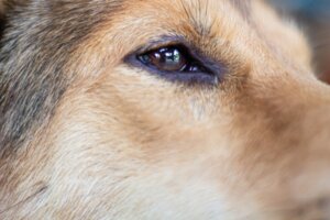
Horner’s syndrome in dogs is a pathology that affects the nerves that connect the eye to the brain, as well as the facial muscles. Generally, it only manifests on one side of the face, but, in rare cases, it can be bilateral.
Although it’s a disease that can occur in any dog breed, Golden Retrievers and Cocker Spaniels have a higher incidence of occurrence. In the following lines, we’ll tell you what causes this ailment, what its most common clinical signs are, and what prevention possibilities exist.
What is Horner’s syndrome?
Horner’s syndrome is actually a group of anomalies that affect the movement of certain facial muscles. This is caused by a malfunction in the nerves that regulate these movements, so that they become erratic and exaggerated.
Causes of Horner’s syndrome in dogs
Horner’s syndrome in dogs appears when some of the nerves that go from the eye to the brain are damaged. Although there are many factors, there are 3 ‘common injuries that can cause the syndrome. We’ll indicate them below.
1. Central lesion (first-order)
In a central injury, the nerve is damaged somewhere before it leaves the spinal cord. The most common causes that provoke this are tumors in the medulla, brain tumors, or trauma in this region. In addition to Horner’s syndrome, other neurological signs may arise, such as motor incoordination or head tilt.
This type of lesion may appear as an effect of some traumas, infarcts, neoplasms, or inflammatory diseases. However, it’s quite rare for these affectations to the nervous system to cause Horner’s syndrome.

2. Preganglionic lesion (second-order)
The damage has been caused in the nerves leading from the medulla to the synapse -the junction between one neuron and another. This lesion arises from neck trauma, tumors in the cervical region, infarcts, or inflammatory processes. It’s also possible for this type of damage to appear as a consequence of thoracic surgeries, strangulation wounds, or bites.
3. Postganglionic lesion (third-order)
The lesion occurs between the synapse and the eye. The most frequent causes of this type of injury are cleaning the dog’s ear with too much force or injury to the animal’s middle ear. However, most postganglionic lesions have an unknown cause.
Most dogs with Horner’s syndrome have postganglionic lesions. In fact, a large proportion of cases are caused by otitis, as neoplasms and injuries in this area of the ear don’t tend to cause problems with the nerves.
Clinical symptoms of Horner’s syndrome in dogs
The clinical symptoms of Horner’s syndrome are very similar to other ocular pathologies. To make a good diagnosis, the intervention of a veterinarian specialized in neurology is absolutely necessary.
The noticeable signs in dogs suffering from the pathology are concentrated in the affected eye and also in the adjacent region in some cases. Some of the most common are as follows:
- Drooping eyelid.
- Contracted pupil or miosis
- Eye sinking or enophthalmos.
- Exposed or prolapsed third eyelid, also known as conjunctival hyperemia.
- Warmer pinna (rare).
Many neurological lesions or the effect of certain drugs can produce these same clinical signs. For this reason, it’s crucial that a neurologist, in addition to the regular veterinarian, examines the pet.
Specialized diagnosis
Any veterinarian can diagnose the syndrome based on clinical signs. However, as these can appear in multiple pathologies, it’s crucial to find out what’s happening at a neurological level and where the lesion has occurred.
In general, it’s estimated that about half of the cases of Horner’s syndrome in dogs are idiopathic in origin. This means that there’s no conclusive cause, as the damage may be due to different pathologies or previous lesions.
The drug used to make the diagnosis is phenylephrine. A few drops are administered in the eye and, if all signs and symptoms disappear, the damage has occurred at the postganglionic level. If the eye doesn’t respond, then other diagnostic tests are necessary.
Chest X-rays, blood tests, other pharmacological tests, and even an MRI may be necessary to find the lesion causing the syndrome. Once the mismatch is found, it’s time to define the course of action and possible treatments.
Treatment and prevention of Horner’s syndrome in dogs
Treatment for the syndrome will depend on where the neurological injury has occurred. Most commonly, treatment isn’t of vital importance, as the body has the capacity to deal with this damage on its own. However, in the case of tumors, the prognosis is reserved and surgical intervention or the administration of chemotherapeutics may be necessary.
When the origin of the nerve damage is due to otitis, a spinal disease, or an inflammatory process, the first thing to do is to control this affectation. Subsequently, drugs are administered to control the symptoms, such as phenylephrine, as well as artificial tears to maintain the lubrication of the eye. Usually, in these situations, the syndrome disappears after about 6 months.
Most cases of dogs suffering from Horner’s syndrome have idiopathic causes. This means that it isn’t known what has caused the syndrome and it simply appears from one day to the next. As you can imagine, this makes treatment very complicated and the prognosis depends on how the dog reacts to the drugs.
Occasionally, the dog may have been bitten in the neck region or suffered a severe ear pull during a scuffle. This can also cause the syndrome. With time and once the injury is healed, the syndrome will disappear. In all cases, regular veterinary visits and good eye and ear health are the best prevention.
Horner’s syndrome in dogs is a pathology that affects the nerves that connect the eye to the brain, as well as the facial muscles. Generally, it only manifests on one side of the face, but, in rare cases, it can be bilateral.
Although it’s a disease that can occur in any dog breed, Golden Retrievers and Cocker Spaniels have a higher incidence of occurrence. In the following lines, we’ll tell you what causes this ailment, what its most common clinical signs are, and what prevention possibilities exist.
What is Horner’s syndrome?
Horner’s syndrome is actually a group of anomalies that affect the movement of certain facial muscles. This is caused by a malfunction in the nerves that regulate these movements, so that they become erratic and exaggerated.
Causes of Horner’s syndrome in dogs
Horner’s syndrome in dogs appears when some of the nerves that go from the eye to the brain are damaged. Although there are many factors, there are 3 ‘common injuries that can cause the syndrome. We’ll indicate them below.
1. Central lesion (first-order)
In a central injury, the nerve is damaged somewhere before it leaves the spinal cord. The most common causes that provoke this are tumors in the medulla, brain tumors, or trauma in this region. In addition to Horner’s syndrome, other neurological signs may arise, such as motor incoordination or head tilt.
This type of lesion may appear as an effect of some traumas, infarcts, neoplasms, or inflammatory diseases. However, it’s quite rare for these affectations to the nervous system to cause Horner’s syndrome.
2. Preganglionic lesion (second-order)
The damage has been caused in the nerves leading from the medulla to the synapse -the junction between one neuron and another. This lesion arises from neck trauma, tumors in the cervical region, infarcts, or inflammatory processes. It’s also possible for this type of damage to appear as a consequence of thoracic surgeries, strangulation wounds, or bites.
3. Postganglionic lesion (third-order)
The lesion occurs between the synapse and the eye. The most frequent causes of this type of injury are cleaning the dog’s ear with too much force or injury to the animal’s middle ear. However, most postganglionic lesions have an unknown cause.
Most dogs with Horner’s syndrome have postganglionic lesions. In fact, a large proportion of cases are caused by otitis, as neoplasms and injuries in this area of the ear don’t tend to cause problems with the nerves.
Clinical symptoms of Horner’s syndrome in dogs
The clinical symptoms of Horner’s syndrome are very similar to other ocular pathologies. To make a good diagnosis, the intervention of a veterinarian specialized in neurology is absolutely necessary.
The noticeable signs in dogs suffering from the pathology are concentrated in the affected eye and also in the adjacent region in some cases. Some of the most common are as follows:
- Drooping eyelid.
- Contracted pupil or miosis
- Eye sinking or enophthalmos.
- Exposed or prolapsed third eyelid, also known as conjunctival hyperemia.
- Warmer pinna (rare).
Many neurological lesions or the effect of certain drugs can produce these same clinical signs. For this reason, it’s crucial that a neurologist, in addition to the regular veterinarian, examines the pet.
Specialized diagnosis
Any veterinarian can diagnose the syndrome based on clinical signs. However, as these can appear in multiple pathologies, it’s crucial to find out what’s happening at a neurological level and where the lesion has occurred.
In general, it’s estimated that about half of the cases of Horner’s syndrome in dogs are idiopathic in origin. This means that there’s no conclusive cause, as the damage may be due to different pathologies or previous lesions.
The drug used to make the diagnosis is phenylephrine. A few drops are administered in the eye and, if all signs and symptoms disappear, the damage has occurred at the postganglionic level. If the eye doesn’t respond, then other diagnostic tests are necessary.
Chest X-rays, blood tests, other pharmacological tests, and even an MRI may be necessary to find the lesion causing the syndrome. Once the mismatch is found, it’s time to define the course of action and possible treatments.
Treatment and prevention of Horner’s syndrome in dogs
Treatment for the syndrome will depend on where the neurological injury has occurred. Most commonly, treatment isn’t of vital importance, as the body has the capacity to deal with this damage on its own. However, in the case of tumors, the prognosis is reserved and surgical intervention or the administration of chemotherapeutics may be necessary.
When the origin of the nerve damage is due to otitis, a spinal disease, or an inflammatory process, the first thing to do is to control this affectation. Subsequently, drugs are administered to control the symptoms, such as phenylephrine, as well as artificial tears to maintain the lubrication of the eye. Usually, in these situations, the syndrome disappears after about 6 months.
Most cases of dogs suffering from Horner’s syndrome have idiopathic causes. This means that it isn’t known what has caused the syndrome and it simply appears from one day to the next. As you can imagine, this makes treatment very complicated and the prognosis depends on how the dog reacts to the drugs.
Occasionally, the dog may have been bitten in the neck region or suffered a severe ear pull during a scuffle. This can also cause the syndrome. With time and once the injury is healed, the syndrome will disappear. In all cases, regular veterinary visits and good eye and ear health are the best prevention.
All cited sources were thoroughly reviewed by our team to ensure their quality, reliability, currency, and validity. The bibliography of this article was considered reliable and of academic or scientific accuracy.
- Boydell, P. (2000). Idiopathic horner syndrome in the golden retriever. Journal of neuro-ophthalmology: the official journal of the North American Neuro-Ophthalmology Society, 20(4), 288-290.
- Herrera, H. D., Suranit, A. P., & Kojusner, N. F. (1998). Idiopathic Horner’s syndrome in collie dogs. Vet Ophthalmol, 1(1), 17-20.
- Morgan, R. V., & Zanotti, S. W. (1989). Horner’s syndrome in dogs and cats: 49 cases (1980-1986). Journal of the American Veterinary Medical Association, 194(8), 1096-1099.
- Simpson, K. M., Williams, D. L., & Cherubini, G. B. (2015). Neuropharmacological lesion localization in idiopathic H orner’s syndrome in Golden Retrievers and dogs of other breeds. Veterinary ophthalmology, 18(1), 1-5.
- Raschia, A., Suarez, S. & Alvarez, M. (2016) Síndrome de Horner producido por linfoma mediastínico en un canino. (Trabajo de grado, UNCPBA).
- Martín, I. (2021) Estudio de los Síndromes de Horner y Key-Gaskell en el perro y en el gato. (Trabajo de Grado, Universidad Zaragoza).
This text is provided for informational purposes only and does not replace consultation with a professional. If in doubt, consult your specialist.