Hyphema in Dogs: What Causes It and How to Treat It?

Written and verified by the veterinarian and zootechnician Sebastian Ramirez Ocampo
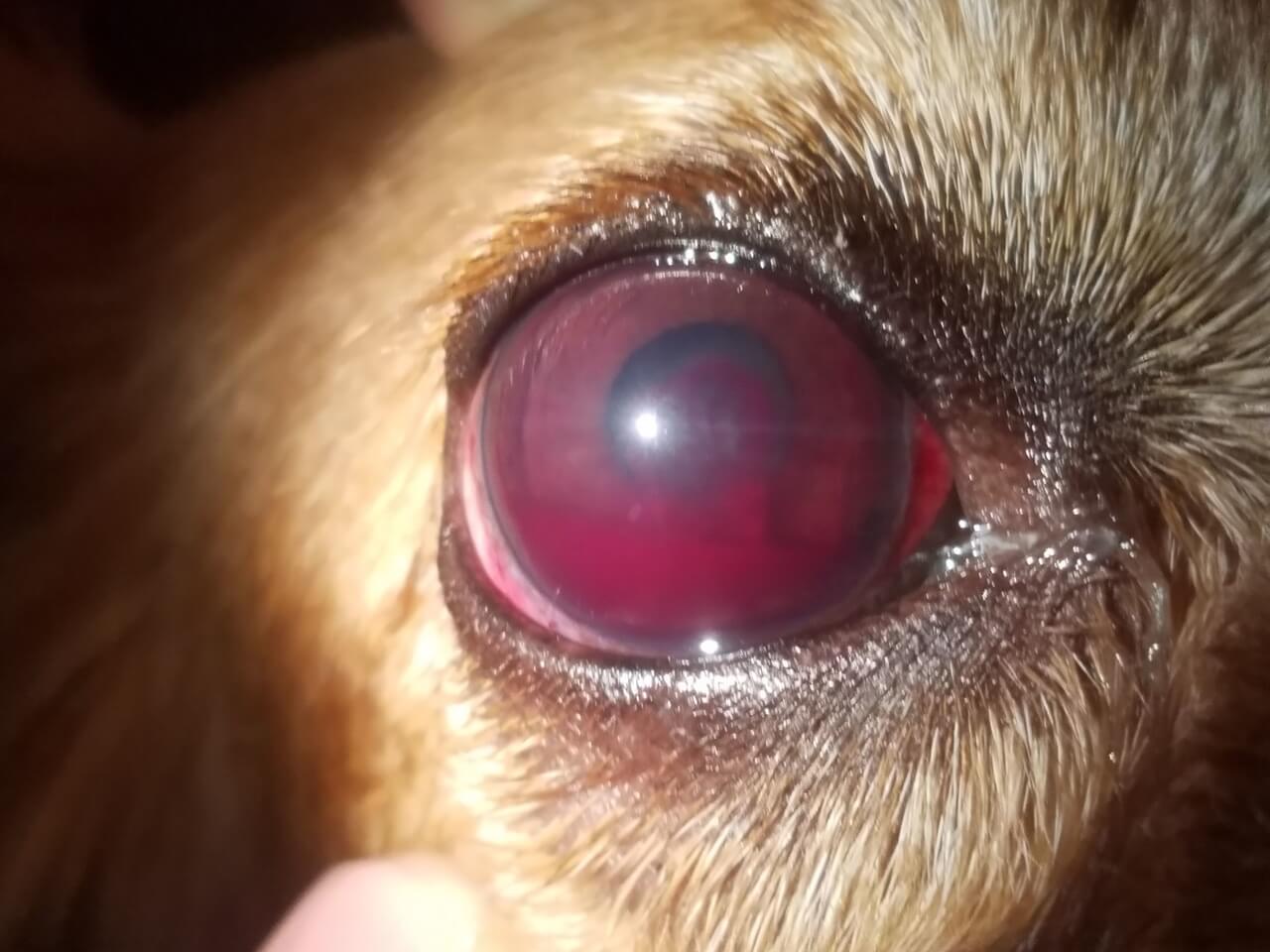
Hyphema in dogs is defined as the presence and accumulation of blood in the eyeball. Among its causes are pathologies of the eye or diseases that affect the body in general.
Due to its implications on animal health, hyphema must be treated urgently. If not, your pet’s vision could be at risk. We’ll tell you all about the causes, complications, diagnosis, and treatment of one of the most frequent ophthalmologic disorders in pets.
What is hyphema in dogs?
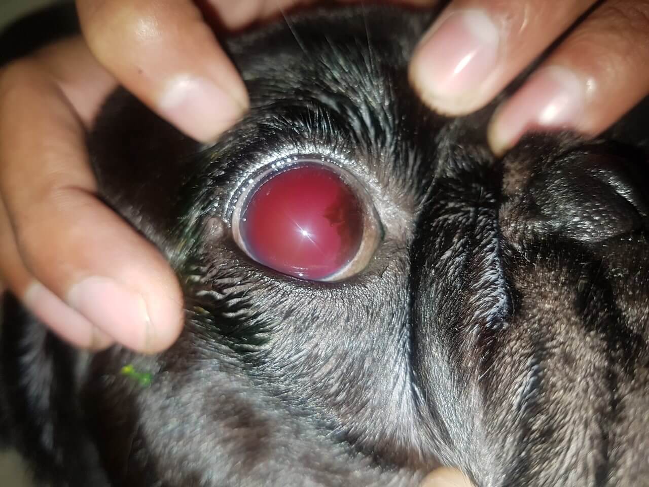
The term “hyphema” refers to the accumulation of blood in the anterior chamber of the eye. This part of the eyeball is the space between the inner surface of the cornea and the surface of the iris. In turn, this compartment is filled with a fluid known as aqueous humor, whose main function is to nourish the anterior structures of the eye.
In general, this pathology occurs after damage and bleeding of the retinal or uveal vasculature, with the consequent mixing of the blood with the vitreous humor: a gelatinous substance that gives volume to the eye and supports the retina. According to the severity and the volume it occupies in the chamber, hyphema is classified as follows:
- Grade I: Blood occupies less than one-third of the anterior chamber.
- Grade II: Blood fills over one-third and up to half of the chamber.
- Grade III: It occupies up to three-quarters of the area.
- Grade IV: The entire anterior chamber is filled with blood.
Due to the effects of gravity, the blood can be observed at the ventral level of the eye. However, when the dog moves suddenly, the blood may fill the chamber again.
Similarly, in these processes, clots aren’t usually present until 4 to 7 days later. This is because the release of fibrinolysins by the iris hinders coagulation. Bleeding may also compromise the drainage pathways of the eye, which may predispose to increased intraocular pressure.
Causes of hyphema in dogs
The presence of blood in your dog’s eye can occur as a result of intraocular conditions or a systemic disease. Severe blunt trauma, uveitis, and coagulation disorders are some of the most common pathologic processes that cause hyphema.
Ocular etiology
The reasons for hyphema of ocular origin are diverse. In most of these cases, the condition occurs in only one eye. The main disorders include the following:
- Trauma: This is listed as the most frequent cause of hyphema in dogs. It may be secondary to cranioencephalic trauma or penetrating ocular wounds, which injure the eyeball or uveal tissues.
- Local tumors: Among the ones most commonly associated with hyphema is intraocular melanoma.
- Uveitis: An inflammation of the uvea or intermediate layer of the eye may cause an extravasation of blood into the anterior chamber.
- Diseases of the retina: Any incident affecting the retina can cause hyphema. This is due to the significant vascularization of this tissue. For this reason, in the event of damage, there may be direct leakage of blood that travels to the anterior chamber.
- Congenital anomalies include disorders such as Collie eye anomaly, persistent hyaloid artery, and vitreoretinal dysplasia.
Systemic etiology
Unlike the above, hyphema caused by systemic diseases is mainly bilateral. In this type of pathology, the following are found:
- Coagulopathies and bleeding disorders: Among the main disorders responsible for hyphema are thrombocytopenia, hemolytic anemia and coagulation problems, for example, as a result of rodenticide poisoning.
- Cancer: The development of hyphema in dogs has been reported in systemic neoplasms such as lymphoma, myeloma, and leukemia.
- Infections: Numerous infections can cause vasculitis and extravasation of blood into the anterior chamber of the eye. The most relevant are ehrlichiosis, brucellosis, and leptospirosis.
- Systemic arterial hypertension: In response to increased pressure in the retinal and choroidal blood vessels, vasoconstriction and increased vascular permeability occurs. This phenomenon can cause ocular problems such as retinal hemorrhages, retinal detachment, and hyphema.
What are the possible complications?
This ocular disorder is considered an emergency, both because of the diseases that can cause it and the complications it’s capable of causing. Among the most serious are the following:
- Formation of intraocular synechiae: If the bleeding progresses towards the formation of clots, adhesions of these to the corneal endothelium or iris may be generated.
- Glaucoma: When these adhesions are created, the iris closes the drainage of aqueous humor, leading to increased intraocular pressure and then glaucoma.
- Blindness: The increase in intraocular pressure, caused by the aqueous humor and bleeding, causes damage to the optic nerve. It leads to blindness, if not treated in time.
- Cataracts have been found as a consequence of chronic hyphema or of chronic or poorly managed hyphemas.
- Phthisis bulbi. If any of the complications or eye diseases that cause hyphema persist over time, the eyeball atrophies or degenerates. This causes it to stop functioning.
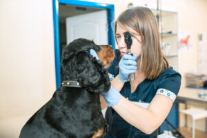
Diagnosis
In essence, the diagnosis is based on identifying the origin or cause of hyphema. Ophthalmological examination is sometimes difficult, because blood doesn’t allow the internal structures of the eye to be seen well.
However, it’s necessary to evaluate certain aspects such as pupillary reflex and intraocular pressure. If possible, the anterior chamber of the eye should also be observed using slit lamps.
Similarly, if your dog suffers from this disorder, the veterinarian may examine the state of blood pressure and recommend certain tests. In this way, he’ll be able to assess the functioning of the organism in general. In addition to this, ocular ultrasound can facilitate the exploration of the eye and bring the definitive diagnosis closer.
In turn, hyphema in dogs must be differentiated from other similar pathologies, such as hemorrhage in the vitreous chamber (which is between the crystalline lens and the back of the eye), hemorrhagic inflammatory exudate, and corneal or iris hemorrhage.
Treatment
As it’s considered an emergency, hyphema should be treated promptly. First of all, the use of steroidal anti-inflammatory drugs topical and systemic, such as dexamethasone and prednisolone.
Corticosteroids of this type prevent the recurrence of bleeding – by inhibiting fibrinolysis – and help to restore the blood-ocular barrier. However, before topical administration, the presence of corneal ulcers should be ruled out. It’s important to do so, because it has been shown that they delay the healing of this membrane.
The use of non-steroidal anti-inflammatory drugs (NSAIDs) isn’t recommended in hyphemas of hemorrhagic origin, because they interfere with platelet function and there may be a higher risk of bleeding. In these cases, they can be replaced by opioid analgesics such as tramadol.
To prevent complications such as synechiae, the topical use of cycloplegic drugs such as atropine or phenylephrine is advised. However, the use of these drugs may increase intraocular pressure. When this phenomenon is observed, they should be discontinued immediately.
Finally, in cases where coagulation is present, the use of tissue plasminogen activator (tPA) is recommended. This fibrinolytic agent, used within 72 hours of clot formation, has been shown to promote rapid clot resolution.
Prognosis
Correct treatment allows this ocular disorder to resolve within 7 to 21 days. Even so, the recovery time will depend on its severity.
When treating grade I hyphemas, recovery occurs in a matter of days, while grade II and III hyphemas can take weeks. In cases where this ocular emergency is classified as grade IV, it’s usually associated with more serious problems, such as ocular atrophy.
According to an article in the journal Veterinary Ophthalmology, the worst prognoses for hyphema are related to severe ocular trauma, neoplasms, and chronic uveitis. In addition, when cataracts or glaucoma are present, the prognosis is guarded.
Hyphema in dogs, a veterinary emergency
As you may have noticed, hyphema is an ophthalmologic emergency that needs quick attention to avoid permanent damage to the canine’s eye. Its manifestation is an indication that our pet’s life is at risk, due to its relationship with serious diseases such as hypertension or poisoning with poisons.
In view of this, at any sign of ocular bleeding, you should immediately go to the veterinarian for them to treat your dog. Remember to perform periodic check-ups and evaluations, so that the vet can identify any aspect that you may have overlooked.
Hyphema in dogs is defined as the presence and accumulation of blood in the eyeball. Among its causes are pathologies of the eye or diseases that affect the body in general.
Due to its implications on animal health, hyphema must be treated urgently. If not, your pet’s vision could be at risk. We’ll tell you all about the causes, complications, diagnosis, and treatment of one of the most frequent ophthalmologic disorders in pets.
What is hyphema in dogs?
The term “hyphema” refers to the accumulation of blood in the anterior chamber of the eye. This part of the eyeball is the space between the inner surface of the cornea and the surface of the iris. In turn, this compartment is filled with a fluid known as aqueous humor, whose main function is to nourish the anterior structures of the eye.
In general, this pathology occurs after damage and bleeding of the retinal or uveal vasculature, with the consequent mixing of the blood with the vitreous humor: a gelatinous substance that gives volume to the eye and supports the retina. According to the severity and the volume it occupies in the chamber, hyphema is classified as follows:
- Grade I: Blood occupies less than one-third of the anterior chamber.
- Grade II: Blood fills over one-third and up to half of the chamber.
- Grade III: It occupies up to three-quarters of the area.
- Grade IV: The entire anterior chamber is filled with blood.
Due to the effects of gravity, the blood can be observed at the ventral level of the eye. However, when the dog moves suddenly, the blood may fill the chamber again.
Similarly, in these processes, clots aren’t usually present until 4 to 7 days later. This is because the release of fibrinolysins by the iris hinders coagulation. Bleeding may also compromise the drainage pathways of the eye, which may predispose to increased intraocular pressure.
Causes of hyphema in dogs
The presence of blood in your dog’s eye can occur as a result of intraocular conditions or a systemic disease. Severe blunt trauma, uveitis, and coagulation disorders are some of the most common pathologic processes that cause hyphema.
Ocular etiology
The reasons for hyphema of ocular origin are diverse. In most of these cases, the condition occurs in only one eye. The main disorders include the following:
- Trauma: This is listed as the most frequent cause of hyphema in dogs. It may be secondary to cranioencephalic trauma or penetrating ocular wounds, which injure the eyeball or uveal tissues.
- Local tumors: Among the ones most commonly associated with hyphema is intraocular melanoma.
- Uveitis: An inflammation of the uvea or intermediate layer of the eye may cause an extravasation of blood into the anterior chamber.
- Diseases of the retina: Any incident affecting the retina can cause hyphema. This is due to the significant vascularization of this tissue. For this reason, in the event of damage, there may be direct leakage of blood that travels to the anterior chamber.
- Congenital anomalies include disorders such as Collie eye anomaly, persistent hyaloid artery, and vitreoretinal dysplasia.
Systemic etiology
Unlike the above, hyphema caused by systemic diseases is mainly bilateral. In this type of pathology, the following are found:
- Coagulopathies and bleeding disorders: Among the main disorders responsible for hyphema are thrombocytopenia, hemolytic anemia and coagulation problems, for example, as a result of rodenticide poisoning.
- Cancer: The development of hyphema in dogs has been reported in systemic neoplasms such as lymphoma, myeloma, and leukemia.
- Infections: Numerous infections can cause vasculitis and extravasation of blood into the anterior chamber of the eye. The most relevant are ehrlichiosis, brucellosis, and leptospirosis.
- Systemic arterial hypertension: In response to increased pressure in the retinal and choroidal blood vessels, vasoconstriction and increased vascular permeability occurs. This phenomenon can cause ocular problems such as retinal hemorrhages, retinal detachment, and hyphema.
What are the possible complications?
This ocular disorder is considered an emergency, both because of the diseases that can cause it and the complications it’s capable of causing. Among the most serious are the following:
- Formation of intraocular synechiae: If the bleeding progresses towards the formation of clots, adhesions of these to the corneal endothelium or iris may be generated.
- Glaucoma: When these adhesions are created, the iris closes the drainage of aqueous humor, leading to increased intraocular pressure and then glaucoma.
- Blindness: The increase in intraocular pressure, caused by the aqueous humor and bleeding, causes damage to the optic nerve. It leads to blindness, if not treated in time.
- Cataracts have been found as a consequence of chronic hyphema or of chronic or poorly managed hyphemas.
- Phthisis bulbi. If any of the complications or eye diseases that cause hyphema persist over time, the eyeball atrophies or degenerates. This causes it to stop functioning.
Diagnosis
In essence, the diagnosis is based on identifying the origin or cause of hyphema. Ophthalmological examination is sometimes difficult, because blood doesn’t allow the internal structures of the eye to be seen well.
However, it’s necessary to evaluate certain aspects such as pupillary reflex and intraocular pressure. If possible, the anterior chamber of the eye should also be observed using slit lamps.
Similarly, if your dog suffers from this disorder, the veterinarian may examine the state of blood pressure and recommend certain tests. In this way, he’ll be able to assess the functioning of the organism in general. In addition to this, ocular ultrasound can facilitate the exploration of the eye and bring the definitive diagnosis closer.
In turn, hyphema in dogs must be differentiated from other similar pathologies, such as hemorrhage in the vitreous chamber (which is between the crystalline lens and the back of the eye), hemorrhagic inflammatory exudate, and corneal or iris hemorrhage.
Treatment
As it’s considered an emergency, hyphema should be treated promptly. First of all, the use of steroidal anti-inflammatory drugs topical and systemic, such as dexamethasone and prednisolone.
Corticosteroids of this type prevent the recurrence of bleeding – by inhibiting fibrinolysis – and help to restore the blood-ocular barrier. However, before topical administration, the presence of corneal ulcers should be ruled out. It’s important to do so, because it has been shown that they delay the healing of this membrane.
The use of non-steroidal anti-inflammatory drugs (NSAIDs) isn’t recommended in hyphemas of hemorrhagic origin, because they interfere with platelet function and there may be a higher risk of bleeding. In these cases, they can be replaced by opioid analgesics such as tramadol.
To prevent complications such as synechiae, the topical use of cycloplegic drugs such as atropine or phenylephrine is advised. However, the use of these drugs may increase intraocular pressure. When this phenomenon is observed, they should be discontinued immediately.
Finally, in cases where coagulation is present, the use of tissue plasminogen activator (tPA) is recommended. This fibrinolytic agent, used within 72 hours of clot formation, has been shown to promote rapid clot resolution.
Prognosis
Correct treatment allows this ocular disorder to resolve within 7 to 21 days. Even so, the recovery time will depend on its severity.
When treating grade I hyphemas, recovery occurs in a matter of days, while grade II and III hyphemas can take weeks. In cases where this ocular emergency is classified as grade IV, it’s usually associated with more serious problems, such as ocular atrophy.
According to an article in the journal Veterinary Ophthalmology, the worst prognoses for hyphema are related to severe ocular trauma, neoplasms, and chronic uveitis. In addition, when cataracts or glaucoma are present, the prognosis is guarded.
Hyphema in dogs, a veterinary emergency
As you may have noticed, hyphema is an ophthalmologic emergency that needs quick attention to avoid permanent damage to the canine’s eye. Its manifestation is an indication that our pet’s life is at risk, due to its relationship with serious diseases such as hypertension or poisoning with poisons.
In view of this, at any sign of ocular bleeding, you should immediately go to the veterinarian for them to treat your dog. Remember to perform periodic check-ups and evaluations, so that the vet can identify any aspect that you may have overlooked.
All cited sources were thoroughly reviewed by our team to ensure their quality, reliability, currency, and validity. The bibliography of this article was considered reliable and of academic or scientific accuracy.
- Book, P., Van der Woerdt, A., & Wilkie, D. (2008). Ultrasonographic abnormalities in eyes with traumatic hyphema obscuring intraocular structures: 33 cases (1991-2002). Journal of Veterinary Emergence and Critical Care, 18 (4). https://onlinelibrary.wiley.com/doi/abs/10.1111/j.1476-4431.2008.00329.x
- Bustamante, L., Rivas, J., & Vargas, P. (2017). Exploración ecográfica ocular básica en perros (modo B tiempo real). Revista de Medicina Veterinaria, 33. http://www.scielo.org.co/scielo.php?script=sci_arttext&pid=S0122-93542017000100113
- Chen, E. J., & Fasiuddin, A. (2021). Management of Traumatic Hyphema and Prevention of Its Complications. Cureus, 13(6), e15771. https://www.ncbi.nlm.nih.gov/pmc/articles/PMC8291460/
- Dehghanpir, S. D., Sheridan, C., Evans, D., Lewin, A. C., Boudreaux, B., & Carter, R. T. (2022). Hyphema and secondary glaucoma as a presenting complaint in a dog with chronic lymphocytic leukemia. Clinical Case Reports, 10(11), e6575. https://www.ncbi.nlm.nih.gov/pmc/articles/PMC9675388/
- Haritha, G., Saritha, G., & Nalini, K. (2016). Clinical Management of Traumatic Hyphema. Journal of Agriculture and Veterinary Science, 9 (9), 11-12. https://www.iosrjournals.org/iosr-javs/papers/vol9-issue9/Version-2/C0909021112.pdf
- Huguet, E., & Delgado, C. (2013). Capítulo 1: Hifema. La oftalmología en colores. Multimédica Ediciones Veterinarias. https://www.berri.es/pdf/OJO%20ROJO‚%20La%20oftalmologia%20en%20colores/9788496344495
- Jinks, M. R., Olea-Popelka, F., & Freeman, K. S. (2018). Causes and outcomes of dogs presenting with hyphema to a referral hospital in Colorado: a retrospective analysis of 99 cases. Veterinary Ophthalmology, 21(2), 160. https://onlinelibrary.wiley.com/doi/10.1111/vop.12491
- Kolb, H. (2005, 1 de mayo). Gross anatomy of the eye. Web vision: The organization of the retina and visual system. Salt Lake City (UT): University of Utah Health Sciences Center. https://www.ncbi.nlm.nih.gov/books/NBK11534/
- Komáromy, A. M., Ramsey, D. T., Brooks, D. E., Ramsey, C. C., Kallberg, M. E., & Andrew, S. E. (2000). Hyphema. Part II. Diagnosis and Treatment. Compendium on Continuing Education for the Practicing Veterinarian, 22 (1), 74-79, 96-. https://repository.upenn.edu/vet_papers/52
- Laminack, E. B., Myrna, K., & Moore, P. A. (2013). Clinical approach to the canine red eye. Today’s Veterinary Practice, 3, 14. https://todaysveterinarypractice.com/ophthalmology/clinical-approach-to-the-canine-red-eye/
- Leiva, M., Naranjo, C., & Peña, M. T. (2005). Ocular signs of canine monocytic ehrlichiosis: a retrospective study in dogs from Barcelona, Spain. Veterinary ophthalmology, 8(6), 387–393. https://onlinelibrary.wiley.com/doi/10.1111/j.1463-5224.2005.00409.x
- Mansoor Lakooraj, H., Ahmadi-Hamedani, M., Dezfoulian, O., & Selk Ghaffari, M. (2018). Multicentric lymphoma in a Rottweiler dog with bilateral ocular involvement: A case report. Veterinary Research Forum: An International Quarterly Journal, 9(3), 285–288. https://www.ncbi.nlm.nih.gov/pmc/articles/PMC6198158/
- Martins, T., & Barros, S. (2015). Red eyes in the necropsy floor: twenty cases of hyphema in dogs and cats. Pesquisa Veterinaria Brasileira, 35 (1) 55-61. https://www.scielo.br/j/pvb/a/TdBhwL7JJqcRQsNdRRKMHcS/?lang=en
- Nelms, S., Nasisse, M., Davidson, M., & Kirschner, S. (1993). Hyphema associated with retinal disease in dogs: 17 cases (1986-1991) Journal of the American Veterinary Association, 202 (8), 1289-1292. https://europepmc.org/article/med/8496090
- Romairone, A. Hipema canino. Diagnóstico veterinario. Consultado el 14 de mayo de 2023. https://www.diagnosticoveterinario.com/hipema-canino/827?utm_content=expand_article
- Stanley, R. (2007). Management of corneal ulcers in small animals. World Small Animal Veterinary Association World Congress Proceedings. https://www.vin.com/apputil/content/defaultadv1.aspx?id=3860707
- Telle, M. R., & Betbeze, C. (2015). Hyphema: Considerations in the Small Animal Patient. Topics in Companion Animal Medicine, 30(3), 97–106. https://www.ncbi.nlm.nih.gov/pmc/articles/PMC7173179/#bib6
- Trbolova, A. (2014). Occurrence and diagnosis of intraocular melanomas in dogs: 6 clinical cases. Medycyna Weterynaryjna, 70(09). http://www.medycynawet.edu.pl/images/stories/pdf/pdf2014/092014/201409578584.pdf
- Wilcock, B. P., & Peiffer, R. L., Jr (1986). Morphology and behavior of primary ocular melanomas in 91 dogs. Veterinary Pathology, 23(4), 418–424. https://journals.sagepub.com/doi/10.1177/030098588602300411
This text is provided for informational purposes only and does not replace consultation with a professional. If in doubt, consult your specialist.