Anisocoria in Dogs: Causes, Diagnosis and Treatment

Written and verified by the veterinarian and zootechnician Sebastian Ramirez Ocampo
In essence, anisocoria in dogs could be defined as asymmetry in pupil size. That is, one pupil is more dilated or contracted than the other. Although it isn’t a pathology in itself, its appearance is related to alterations in the ocular, nervous, and auditory systems.
For this reason, it’s important for you to know the origin of anisocoria in your pet, in order to provide timely care. Discover all the aspects related to this unusual pupillary disorder.
Pupillary dilation and contraction
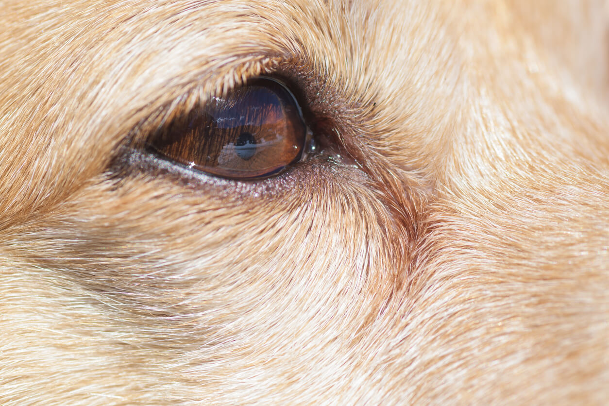
The pupil is the black structure found in the center of the iris. In dogs, its shape is always round, and it’s similar to the diaphragm opening of a camera. It has the function of regulating the entrance of light into the eye, especially into the retina. It does this through two processes known as pupillary dilation and contraction.
The first occurs when there’s a very poor light stimulus. In response, the sympathetic nervous system (SNS) is responsible for increasing the size of the pupil in order to obtain as much light as possible. This is known as mydriasis.
The opposite occurs in pupillary constriction. When there are too many light stimuli, the parasympathetic nervous system (PNS) is responsible for closing the pupil to reduce the entry of light into the pupil. This phenomenon is called miosis.
Causes of anisocoria in dogs
Associate professor at the University of Arizona College of Veterinary Medicine, Ryane Englar, states in her book Common Clinical Presentations in Dogs and Cats published in 2019, that “anisocoria may be a result of primary ocular disease or primary neurologic disease.”
This means that the difference in pupil size in your dog may be a manifestation of some disorder that may occur in the visual or nervous systems. Let’s review the most common causes.
Ocular causes
Anisocoria can be a condition secondary to diseases of the eye or dysfunctions in the optic nerve, which is responsible for transmitting visual stimuli from the retina to the brain.
Asymmetry in pupil size can also appear as a result of the dog’s eye contact with certain plants and substances. When this is the case, we refer to ophthalmologic or ocular causes. These include the following:
- Medications: Atropine and tropicamide cause mydriasis in the pupil, while pilocarpine causes miosis.
- Bromide toxicosis: The administration of potassium bromide, combined with phenobarbital, is common to treat idiopathic epilepsy. However, its excess can cause a toxicosis, known as bromismus, and this in turn is likely to cause anisocoria.
- Exposure to toxic plants: There are certain compounds or biological materials that can cause eye irritation and anisocoria in dogs when direct eye contact occurs. Stramonium (Data stramonium) is a plant that causes this eye disorder in canines.
- Anterior uveitis: This is the inflammation of the iris – where the pupil is located – and part of the middle layer of the ocular wall, called the ciliary body. This affects the pupillary sphincter, causing mydriasis as the pupil cannot contract.
- Hypoplasia of the iris: This is a condition in which the iris doesn’t develop optimally or is very thin. In dogs, it can be congenital or secondary to inflammation of the iris.
- Iris atrophy: This degeneration is associated with the aging of the dog and causes mydriasis.
- Posterior synechiae: When the iris adheres to the lens capsule, the aqueous humor is blocked. The pupil may be abnormally shaped in the affected eye.
- Glaucoma: This consists of the increase of the intraocular pressure, a product of the accumulation of aqueous humor. The progression of this disease causes damage to the optic nerve, with the consequent loss of vision. Likewise, it can affect the iris and pupil, causing mydriasis in the affected eye.
Neurological causes
Asymmetry in pupil size may also be a manifestation of some dysfunction in the sympathetic or parasympathetic systems. Neurological causes include afferent or sensory lesions and efferent or motor lesions. In companion animals, the following are most frequently present:
- Craniocerebral trauma: A cranial trauma can create a serious injury or inflammation in the nerves that communicate the nervous system with the eye.
- Intracranial neoplasia: A tumor in the central nervous system is capable of causing interference in the communication of the brain with the nerve pathways that innervate the pupil.
- Organophosphate toxicity: These are ingredients present in insecticides and herbicides. When ingested, they produce intoxication manifested by signs such as excessive salivation, lacrimation, urination, and in some cases, anisocoria.
- Sympathetic denervation or Horner’s syndrome: This occurs when the sympathetic pathway, responsible for pupil dilation, is interrupted. It’s characterized by miosis of the affected pupil and protrusion or displacement of the third eyelid.
- Parasympathetic denervation: An alteration in the parasympathetic pathway prevents pupillary contraction.
- Concurrent sympathetic and parasympathetic denervation or dysautonomia: This is the simultaneous presence of both dysfunctions. Although it’s more common in cats than in dogs, it tends to manifest among medium to large canines living in rural areas.
Other causes
Otitis media can cause anisocoria. The neurons and nerves involved in the communication of the pupil with the brain pass through the auditory canal. For this reason, an inflammation of the middle ear can generate interference in this nerve pathway. In this case, miosis or persistent contraction of the pupil occurs.
On the other hand, some tick bites can transmit canine ehrlichiosis. According to an article published in the journal Veterinary Sciences, anisocoria has been observed among the ocular signs shown by some canines with this infectious disease.
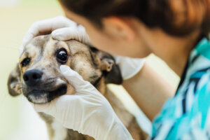
Diagnosing the cause of anisocoria in dogs
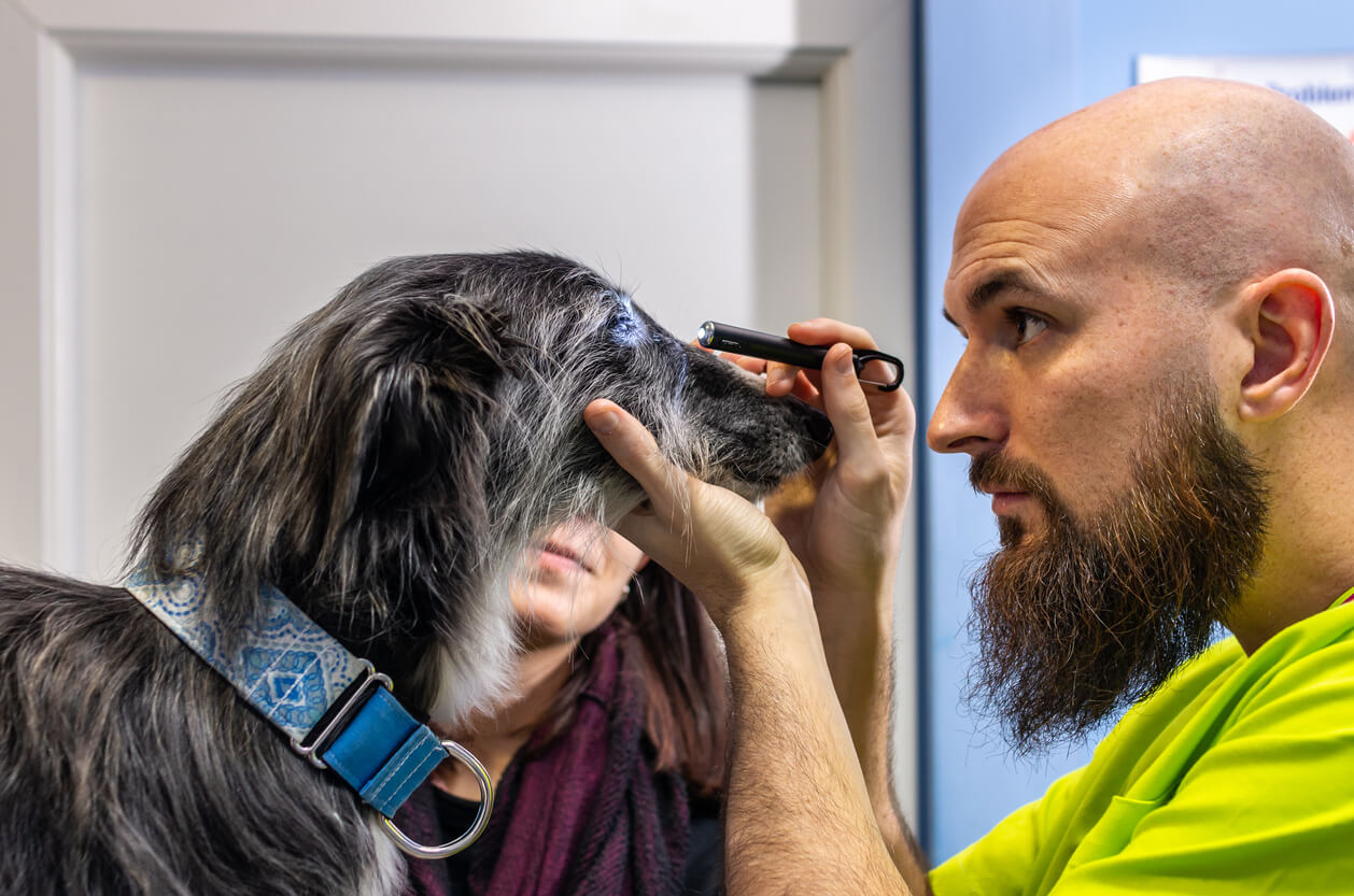
The location of the lesion is essential to be able to confirm the diagnosis and treatment of the condition underlying the asymmetry in pupil size. To do this, the veterinarian will perform a physical examination, including an eye exam, to identify which of the two pupils is affected. To do this, the canine is evaluated in bright, dim light.
In addition to this, other tests may be performed. These include the Schirmer’s test that measures tear production. In addition, a fluorescein test can determine if there are foreign bodies or an ulcer in the cornea. It consists of administering an orange dye on the ocular surface and illuminating the eye with a cobalt blue light.
Tonometry is a procedure performed to measure the intraocular pressure in the dog’s eyes. This evaluation allows a vet to detect if they have glaucoma or uveitis.
The specialist can also perform pharmacological tests – with physostigmine, diluted pilocarpine or diluted phenylephrine – in order to distinguish if the dysfunction is in the sympathetic or parasympathetic pathways. Similarly, they may recommend blood and urine tests, as well as a CT scan or an MRI.
Management and final considerations of anisocoria in dogs
As you can see, anisocoria in dogs has different origins. From trauma, tumors, nervous system problems to inflammation of the ear and the eye itself. In these cases, the veterinarian will be in charge of identifying what is causing this problem in your pet, in order to provide the appropriate treatment.
In general, diseases that cause anisocoria can be treated medically, so there’s no need to panic if you observe this symptom in your dog. However, it’s your duty to identify it quickly, because timely attention will always make a difference.
In essence, anisocoria in dogs could be defined as asymmetry in pupil size. That is, one pupil is more dilated or contracted than the other. Although it isn’t a pathology in itself, its appearance is related to alterations in the ocular, nervous, and auditory systems.
For this reason, it’s important for you to know the origin of anisocoria in your pet, in order to provide timely care. Discover all the aspects related to this unusual pupillary disorder.
Pupillary dilation and contraction
The pupil is the black structure found in the center of the iris. In dogs, its shape is always round, and it’s similar to the diaphragm opening of a camera. It has the function of regulating the entrance of light into the eye, especially into the retina. It does this through two processes known as pupillary dilation and contraction.
The first occurs when there’s a very poor light stimulus. In response, the sympathetic nervous system (SNS) is responsible for increasing the size of the pupil in order to obtain as much light as possible. This is known as mydriasis.
The opposite occurs in pupillary constriction. When there are too many light stimuli, the parasympathetic nervous system (PNS) is responsible for closing the pupil to reduce the entry of light into the pupil. This phenomenon is called miosis.
Causes of anisocoria in dogs
Associate professor at the University of Arizona College of Veterinary Medicine, Ryane Englar, states in her book Common Clinical Presentations in Dogs and Cats published in 2019, that “anisocoria may be a result of primary ocular disease or primary neurologic disease.”
This means that the difference in pupil size in your dog may be a manifestation of some disorder that may occur in the visual or nervous systems. Let’s review the most common causes.
Ocular causes
Anisocoria can be a condition secondary to diseases of the eye or dysfunctions in the optic nerve, which is responsible for transmitting visual stimuli from the retina to the brain.
Asymmetry in pupil size can also appear as a result of the dog’s eye contact with certain plants and substances. When this is the case, we refer to ophthalmologic or ocular causes. These include the following:
- Medications: Atropine and tropicamide cause mydriasis in the pupil, while pilocarpine causes miosis.
- Bromide toxicosis: The administration of potassium bromide, combined with phenobarbital, is common to treat idiopathic epilepsy. However, its excess can cause a toxicosis, known as bromismus, and this in turn is likely to cause anisocoria.
- Exposure to toxic plants: There are certain compounds or biological materials that can cause eye irritation and anisocoria in dogs when direct eye contact occurs. Stramonium (Data stramonium) is a plant that causes this eye disorder in canines.
- Anterior uveitis: This is the inflammation of the iris – where the pupil is located – and part of the middle layer of the ocular wall, called the ciliary body. This affects the pupillary sphincter, causing mydriasis as the pupil cannot contract.
- Hypoplasia of the iris: This is a condition in which the iris doesn’t develop optimally or is very thin. In dogs, it can be congenital or secondary to inflammation of the iris.
- Iris atrophy: This degeneration is associated with the aging of the dog and causes mydriasis.
- Posterior synechiae: When the iris adheres to the lens capsule, the aqueous humor is blocked. The pupil may be abnormally shaped in the affected eye.
- Glaucoma: This consists of the increase of the intraocular pressure, a product of the accumulation of aqueous humor. The progression of this disease causes damage to the optic nerve, with the consequent loss of vision. Likewise, it can affect the iris and pupil, causing mydriasis in the affected eye.
Neurological causes
Asymmetry in pupil size may also be a manifestation of some dysfunction in the sympathetic or parasympathetic systems. Neurological causes include afferent or sensory lesions and efferent or motor lesions. In companion animals, the following are most frequently present:
- Craniocerebral trauma: A cranial trauma can create a serious injury or inflammation in the nerves that communicate the nervous system with the eye.
- Intracranial neoplasia: A tumor in the central nervous system is capable of causing interference in the communication of the brain with the nerve pathways that innervate the pupil.
- Organophosphate toxicity: These are ingredients present in insecticides and herbicides. When ingested, they produce intoxication manifested by signs such as excessive salivation, lacrimation, urination, and in some cases, anisocoria.
- Sympathetic denervation or Horner’s syndrome: This occurs when the sympathetic pathway, responsible for pupil dilation, is interrupted. It’s characterized by miosis of the affected pupil and protrusion or displacement of the third eyelid.
- Parasympathetic denervation: An alteration in the parasympathetic pathway prevents pupillary contraction.
- Concurrent sympathetic and parasympathetic denervation or dysautonomia: This is the simultaneous presence of both dysfunctions. Although it’s more common in cats than in dogs, it tends to manifest among medium to large canines living in rural areas.
Other causes
Otitis media can cause anisocoria. The neurons and nerves involved in the communication of the pupil with the brain pass through the auditory canal. For this reason, an inflammation of the middle ear can generate interference in this nerve pathway. In this case, miosis or persistent contraction of the pupil occurs.
On the other hand, some tick bites can transmit canine ehrlichiosis. According to an article published in the journal Veterinary Sciences, anisocoria has been observed among the ocular signs shown by some canines with this infectious disease.
Diagnosing the cause of anisocoria in dogs
The location of the lesion is essential to be able to confirm the diagnosis and treatment of the condition underlying the asymmetry in pupil size. To do this, the veterinarian will perform a physical examination, including an eye exam, to identify which of the two pupils is affected. To do this, the canine is evaluated in bright, dim light.
In addition to this, other tests may be performed. These include the Schirmer’s test that measures tear production. In addition, a fluorescein test can determine if there are foreign bodies or an ulcer in the cornea. It consists of administering an orange dye on the ocular surface and illuminating the eye with a cobalt blue light.
Tonometry is a procedure performed to measure the intraocular pressure in the dog’s eyes. This evaluation allows a vet to detect if they have glaucoma or uveitis.
The specialist can also perform pharmacological tests – with physostigmine, diluted pilocarpine or diluted phenylephrine – in order to distinguish if the dysfunction is in the sympathetic or parasympathetic pathways. Similarly, they may recommend blood and urine tests, as well as a CT scan or an MRI.
Management and final considerations of anisocoria in dogs
As you can see, anisocoria in dogs has different origins. From trauma, tumors, nervous system problems to inflammation of the ear and the eye itself. In these cases, the veterinarian will be in charge of identifying what is causing this problem in your pet, in order to provide the appropriate treatment.
In general, diseases that cause anisocoria can be treated medically, so there’s no need to panic if you observe this symptom in your dog. However, it’s your duty to identify it quickly, because timely attention will always make a difference.
All cited sources were thoroughly reviewed by our team to ensure their quality, reliability, currency, and validity. The bibliography of this article was considered reliable and of academic or scientific accuracy.
- Barnes, H., & Bentley E. (2015, 18 de diciembre). The Practitioner´s Guido To Neurologic Causes of Canine Anisocoria. Today’s Veterinary Practice. https://todaysveterinarypractice.com/ophthalmology/the-practitioners-guide-neurologic-causes-canine-anisocoria/
- Bruyette, D. (Ed.). (2020). Clinical Small Animal Internal Medicine. John Wiley & Sons.
- Clarke, K. E., Sorrell, S., & Breheny, C. (2020). Dysautonomia in 53 cats and dogs: retrospective review of clinical data and outcome. 187(12), e118. https://bvajournals.onlinelibrary.wiley.com/doi/abs/10.1136/vr.105258
- De Lahunta, A., Glass, E. N., & Kent, M. (2020). De Lahunta’s Veterinary Neuroanatomy and Clinical Neurology-E-Book. Elsevier Health Sciences.
- Englar, R. (2019). Common Clinical Presentations in Dogs and Cats. Anisocoria (pp. 271-282). https://www.researchgate.net/publication/335607435_Anisocoria
- Grahn, B. H., Taylor, S. M., & Sandmeyer, L. S. (2007). Diagnostic ophthalmology . Left internal ophthalmoplegia. Can Vet J, 48(3), 321-322. https://www.ncbi.nlm.nih.gov/pmc/articles/PMC1800946/
- Hansen, P., & Clerc, B. (2002). Anisocoria in the dog provoked by a toxic contact with an ornamental plant: Datura stramonium. Vet Ophthalmol, 5(4), 277-279. https://onlinelibrary.wiley.com/doi/abs/10.1046/j.1463-5224.2002.00224.x?sid=nlm%3Apubmed
- Kim, J., Heo, J., Ji, D., & Kim, M. S. (2015). Quantitative assessment of pupillary light reflex in normal and anesthetized dogs: a preliminary study. The Journal of Veterinary Medical Science, 77(4), 475–478. https://www.ncbi.nlm.nih.gov/pmc/articles/PMC4427750/
- Kucera, T. (2023). Anisocoria in Dogs: Causes, Treatment, and Prevention. The Spruce Pets. Consultado el 5 de mayo de 2023. https://www.thesprucepets.com/dog-pupil-sizes-are-unequal-4801666
- Saito, T. B., & Walker, D. H. (2016). Ehrlichioses: an important one health opportunity. Veterinary Sciences, 3(3), 20. https://www.mdpi.com/2306-7381/3/3/20
- Thompson, M. (2017). Small Animal Medical Differential Diagnosis: A Book of Lists. Elsevier Health Sciences.
- Wegg, M. L. (2020). A retrospective evaluation of systemic and/or topical pilocarpine treatment for canine neurogenic dry eye: 11 cases. Veterinary Ophthalmology, 23(2), 341-346. https://doi.org/10.1111/vop.12731
- Yohn, S. E., Morrison, W. B., & Sharp, P. E. (1992). Bromide toxicosis (bromism) in a dog treated with potassium bromide for refractory seizures. Journal of the American Veterinary Medical Association, 201(3), 468–470.
- Zwueste, D. M., & Grahn, B. H. (2019). A review of Horner’s syndrome in small animals. The Canadian Veterinary Journal, 60(1), 81-88. https://www.ncbi.nlm.nih.gov/pmc/articles/PMC6294019/
This text is provided for informational purposes only and does not replace consultation with a professional. If in doubt, consult your specialist.