Corneal Ulcers in Cats: Causes, Types and Treatment

Written and verified by the veterinarian and zootechnician Sebastian Ramirez Ocampo
Corneal ulcers in cats are one of the most frequent ocular pathologies in veterinary practice. In fact, research published in the Revista de Investigaciones Veterinarias del Perú during the years 2014 to 2020, exposed that this type of lesion is the most reported in this species, only followed by conjunctival diseases.
Taking into account the importance of the visual organs, it’s essential that feline owners know the main causes, symptoms, types, and treatment of corneal ulcers. This is in order to take care of your pet in a timely manner. Continue reading and learn all the aspects related to this eye disease.
What are corneal ulcers and what are their causes?
The cornea is an avascular and transparent structure whose main function is to prevent the entry of foreign bodies, microorganisms, and chemical agents into the eye. It also plays a fundamental role in vision.
It’s in charge of controlling the focus and the entrance of light to the eye in order to see images clearly. It’s composed of an outer epithelial layer, the stroma, Descemet’s membrane, and an inner endothelial layer.
In clinical terms, an ulcer could be defined as the loss of continuity of one or more corneal layers, which alters the integrity and functionality of this protective membrane. In general, patients with this condition show signs of pain and ocular discomfort, in addition to a marked opacity of the cornea, according to a study published in the Electronic Journal of Veterinary Medicine.
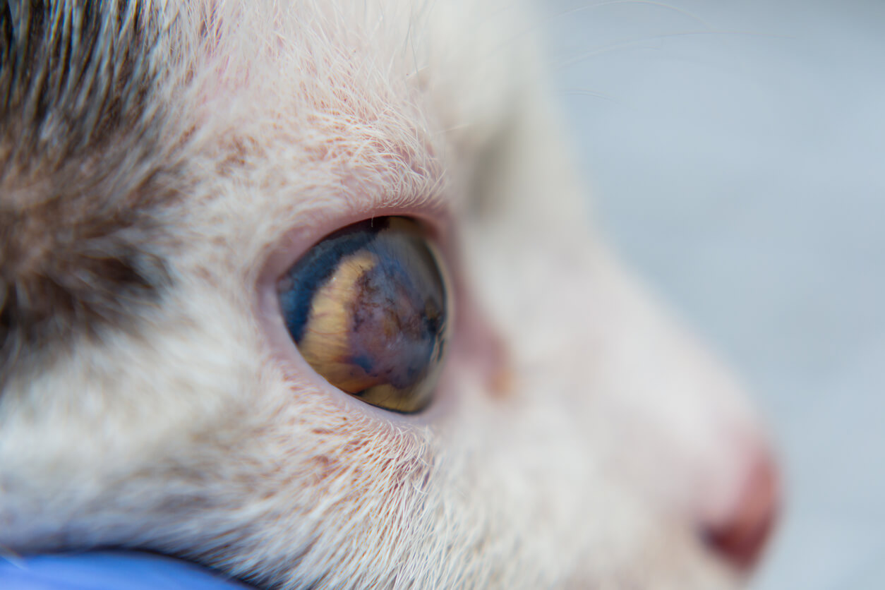
Among the factors that predispose to the formation of corneal ulcers in cats are protruding eyes, access to the exterior, and coexistence with other felines. Likewise, the above study indicates that the main causes for the development of this ocular pathology in cats are the following:
- Traumatic injuries: As their name indicates, these are generated by blows, scratches, and contact of the cornea with foreign bodies or chemical agents.
- Viral diseases: One of the main causes of cat eye ulcers is feline herpesvirus type 1 infection.
- Associated ocular pathologies: Ocular diseases such as keratoconjunctivitis sicca or dry eye, distichiasis, and ectopic cilium, all produce a predisposition to develop corneal ulcers.
Types of corneal ulcers in cats
According to the depth, evolution, and extent of the lesion, ulcers can be classified as simple or complicated:
Simple ulcers
Simple ulcers are lesions that only affect the external corneal epithelium and the surface of the stroma. They’re usually caused by foreign bodies, trauma, dry eye, chemical irritants, viral infections such as herpesvirus-1, and surgical procedures where the eye isn’t properly lubricated.
This type of ulcers are very painful and affected animals show the following symptoms:
- Blepharospasm
- Excessive tearing
- Pupillary contraction
- Sensitivity to light
- Corneal edema
The diagnosis of superficial simple ulcers is made by topical application of dyes such as fluorescein. Likewise, a complete bilateral ophthalmologic examination can be performed. In addition, complementary examinations such as Schirmer’s test, which evaluates the production of tears in the eye, can be added.
Complicated ulcers
Complicated ulcers are those that show abnormalities in healing, infectious processes, the presence of cellular infiltrate, or those that compromise more than half of the corneal structures. Accordingly, these types of lesions include infected ulcers, indolent ulcers, descemetoceles, and perforated lesions. The following will explain what each one consists of.
Infected ulcers
These occur when a corneal lesion becomes contaminated with opportunistic agents, usually bacteria. According to a study published in Veterinary Clinics of North America: Small Animal Practice, these microorganisms can create an increase in the breadth and depth of the lesion, since some bacteria such as Pseudomonas secrete proteolytic enzymes that affect corneal structures.
To identify the infecting microorganism, methods such as cytology, cultures, and antibiograms of samples taken from corneal tissue are used.
Indolent ulcers
These are identified as lesions in which there’s epithelial growth, but without adhesion of the epithelium to the stroma or basement membrane of the epithelium. That is to say, there’s a healing process, but it isn’t fully completed. Therefore, they’re lesions that constantly recur. In cats, they’re associated with feline herpesvirus type 1 infection.
During the acute phase, signs such as blepharospasm, excessive lacrimation, and sensitivity to light may be present. However, as the lesion becomes chronic, patients show no signs of pain or discomfort.
Descemetocele
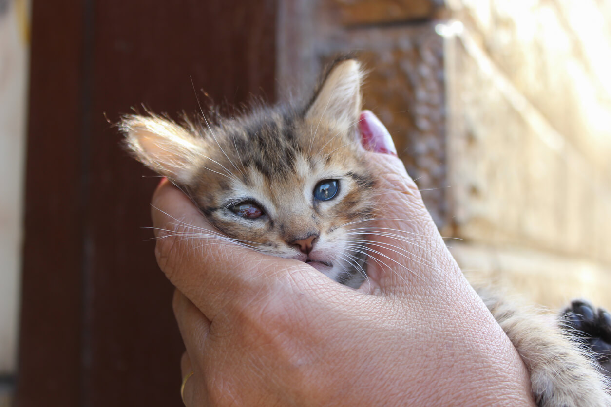
When the ulcer has destroyed the epithelial layer and the stroma has reached Descemet’s membrane, a severe lesion that compromises the integrity of the eye known as a “descemetocele” occurs. This type of ulcer is considered a medical emergency as there’s a risk of ocular perforation. Signs of severe uveitis, cellular infiltrate, and little or no pain symptoms may be present.
Perforated lesions
Perforated ulcers result from trauma, descemetoceles, severe infections, or corneal sequestration in felines. Clinical signs of this injury include blepharospasm, purulent or serous ocular discharge, corneal edema, collapsed anterior chamber, and iris prolapse, with subsequent loss of the eye if prompt treatment isn’t performed.
Treatment of corneal ulcers in cats
Treatment of these ocular pathologies should be based on the severity, complexity, and extent of the lesion. On the one hand, simple or superficial ulcers are treated with antibiotic preparations, anti-inflammatories, and topical analgesics.
It’s important to note that the use of corticosteroid medications is contraindicated, as they interfere with the corneal healing process and its defense mechanisms.
On the other hand, more complicated ulcers are usually treated surgically or with other types of medications. For example, techniques such as grid keratotomy, diamond burr debridement, conjunctival flap, or autologous membrane implants have worked well in the treatment of these cases.
In addition, the use of platelet-rich plasma and the application of stem cells have shown positive results in the resolution of feline eye ulcers.
A disease that deserves attention
As you have seen, corneal ulcers aren’t a pathology to be taken lightly. It’s essential that you identify any sign of eye pain in your pet promptly, in order to treat the cornea in time and prevent it from progressing to deeper or more complicated lesions.
Remember to pay special attention if your cat goes outdoors, since fights with other animals usually cause this type of disease. Try to visit your veterinarian regularly to keep your feline healthy.
Corneal ulcers in cats are one of the most frequent ocular pathologies in veterinary practice. In fact, research published in the Revista de Investigaciones Veterinarias del Perú during the years 2014 to 2020, exposed that this type of lesion is the most reported in this species, only followed by conjunctival diseases.
Taking into account the importance of the visual organs, it’s essential that feline owners know the main causes, symptoms, types, and treatment of corneal ulcers. This is in order to take care of your pet in a timely manner. Continue reading and learn all the aspects related to this eye disease.
What are corneal ulcers and what are their causes?
The cornea is an avascular and transparent structure whose main function is to prevent the entry of foreign bodies, microorganisms, and chemical agents into the eye. It also plays a fundamental role in vision.
It’s in charge of controlling the focus and the entrance of light to the eye in order to see images clearly. It’s composed of an outer epithelial layer, the stroma, Descemet’s membrane, and an inner endothelial layer.
In clinical terms, an ulcer could be defined as the loss of continuity of one or more corneal layers, which alters the integrity and functionality of this protective membrane. In general, patients with this condition show signs of pain and ocular discomfort, in addition to a marked opacity of the cornea, according to a study published in the Electronic Journal of Veterinary Medicine.

Among the factors that predispose to the formation of corneal ulcers in cats are protruding eyes, access to the exterior, and coexistence with other felines. Likewise, the above study indicates that the main causes for the development of this ocular pathology in cats are the following:
- Traumatic injuries: As their name indicates, these are generated by blows, scratches, and contact of the cornea with foreign bodies or chemical agents.
- Viral diseases: One of the main causes of cat eye ulcers is feline herpesvirus type 1 infection.
- Associated ocular pathologies: Ocular diseases such as keratoconjunctivitis sicca or dry eye, distichiasis, and ectopic cilium, all produce a predisposition to develop corneal ulcers.
Types of corneal ulcers in cats
According to the depth, evolution, and extent of the lesion, ulcers can be classified as simple or complicated:
Simple ulcers
Simple ulcers are lesions that only affect the external corneal epithelium and the surface of the stroma. They’re usually caused by foreign bodies, trauma, dry eye, chemical irritants, viral infections such as herpesvirus-1, and surgical procedures where the eye isn’t properly lubricated.
This type of ulcers are very painful and affected animals show the following symptoms:
- Blepharospasm
- Excessive tearing
- Pupillary contraction
- Sensitivity to light
- Corneal edema
The diagnosis of superficial simple ulcers is made by topical application of dyes such as fluorescein. Likewise, a complete bilateral ophthalmologic examination can be performed. In addition, complementary examinations such as Schirmer’s test, which evaluates the production of tears in the eye, can be added.
Complicated ulcers
Complicated ulcers are those that show abnormalities in healing, infectious processes, the presence of cellular infiltrate, or those that compromise more than half of the corneal structures. Accordingly, these types of lesions include infected ulcers, indolent ulcers, descemetoceles, and perforated lesions. The following will explain what each one consists of.
Infected ulcers
These occur when a corneal lesion becomes contaminated with opportunistic agents, usually bacteria. According to a study published in Veterinary Clinics of North America: Small Animal Practice, these microorganisms can create an increase in the breadth and depth of the lesion, since some bacteria such as Pseudomonas secrete proteolytic enzymes that affect corneal structures.
To identify the infecting microorganism, methods such as cytology, cultures, and antibiograms of samples taken from corneal tissue are used.
Indolent ulcers
These are identified as lesions in which there’s epithelial growth, but without adhesion of the epithelium to the stroma or basement membrane of the epithelium. That is to say, there’s a healing process, but it isn’t fully completed. Therefore, they’re lesions that constantly recur. In cats, they’re associated with feline herpesvirus type 1 infection.
During the acute phase, signs such as blepharospasm, excessive lacrimation, and sensitivity to light may be present. However, as the lesion becomes chronic, patients show no signs of pain or discomfort.

Descemetocele
When the ulcer has destroyed the epithelial layer and the stroma has reached Descemet’s membrane, a severe lesion that compromises the integrity of the eye known as a “descemetocele” occurs. This type of ulcer is considered a medical emergency as there’s a risk of ocular perforation. Signs of severe uveitis, cellular infiltrate, and little or no pain symptoms may be present.
Perforated lesions
Perforated ulcers result from trauma, descemetoceles, severe infections, or corneal sequestration in felines. Clinical signs of this injury include blepharospasm, purulent or serous ocular discharge, corneal edema, collapsed anterior chamber, and iris prolapse, with subsequent loss of the eye if prompt treatment isn’t performed.
Treatment of corneal ulcers in cats
Treatment of these ocular pathologies should be based on the severity, complexity, and extent of the lesion. On the one hand, simple or superficial ulcers are treated with antibiotic preparations, anti-inflammatories, and topical analgesics.
It’s important to note that the use of corticosteroid medications is contraindicated, as they interfere with the corneal healing process and its defense mechanisms.
On the other hand, more complicated ulcers are usually treated surgically or with other types of medications. For example, techniques such as grid keratotomy, diamond burr debridement, conjunctival flap, or autologous membrane implants have worked well in the treatment of these cases.
In addition, the use of platelet-rich plasma and the application of stem cells have shown positive results in the resolution of feline eye ulcers.
A disease that deserves attention
As you have seen, corneal ulcers aren’t a pathology to be taken lightly. It’s essential that you identify any sign of eye pain in your pet promptly, in order to treat the cornea in time and prevent it from progressing to deeper or more complicated lesions.
Remember to pay special attention if your cat goes outdoors, since fights with other animals usually cause this type of disease. Try to visit your veterinarian regularly to keep your feline healthy.
All cited sources were thoroughly reviewed by our team to ensure their quality, reliability, currency, and validity. The bibliography of this article was considered reliable and of academic or scientific accuracy.
- Anastassiadis, Z., Bayley, K., & Read, A. (2022). Corneal diamond burr debridement for superficial non-healing corneal ulcers in cats. Veterinary ophthalmology, 25(6), 476-482. https://onlinelibrary.wiley.com/doi/10.1111/vop.13026
- Baraboglia, D. (2009). Uso de la fluoresceína en la practica clínica veterinaria. Revista electrónica de veterinaria, 10 (3). https://www.redalyc.org/pdf/636/63617318012.pdf
- Farghali, H. A., AbdElKader, N. A., AbuBakr, H. O., Ramadan, E. S., Khattab, M. S., Salem, N. Y., & Emam, I. A. (2021). Corneal Ulcer in Dogs and Cats: Novel Clinical Application of Regenerative Therapy Using Subconjunctival Injection of Autologous Platelet-Rich Plasma. Frontiers in veterinary science, 8, 641265. https://doi.org/10.3389/fvets.2021.641265
- Giuliano E. A. (2004). Nonsteroidal anti-inflammatory drugs in veterinary ophthalmology. The Veterinary clinics of North America. Small animal practice, 34(3), 707–723. https://www.sciencedirect.com/science/article/abs/pii/S0195561603001815?via%3Dihub
- Gould, D. (2011). Feline Herpesvirus-1: Ocular manifestations, Diagnosis and Treatment options. Jorunal of Feline Medicine and Surgery, 13 (5). https://journals.sagepub.com/doi/10.1016/j.jfms.2011.03.010?url_ver=Z39.88-2003&rfr_id=ori:rid:crossref.org&rfr_dat=cr_pub%20%200pubmed
- Hartley C. (2010). Treatment of corneal ulcers: what are the medical options?. Journal of feline medicine and surgery, 12(5), 384–397. https://journals.sagepub.com/doi/10.1016/j.jfms.2010.03.012?url_ver=Z39.88-2003&rfr_id=ori:rid:crossref.org&rfr_dat=cr_pub%20%200pubmed
- Hugues, B., & Torres, M. (2022). Enfermedades del sistema ocular diagnosticadas en perros y gatos de La Habana, Cuba. Periodo 2014-2020. Revista de Investigaciones Veterinarias del Perú, 33. http://www.scielo.org.pe/scielo.php?pid=S1609-91172022000200013&script=sci_arttext
- Jégou, J. P., & Tromeur, F. (2015). Superficial keratectomy for chronic corneal ulcers refractory to medical treatment in 36 cats. Veterinary ophthalmology, 18(4), 335–340. https://onlinelibrary.wiley.com/doi/10.1111/vop.12153
- La Croix, N. C., van der Woerdt, A., & Olivero, D. K. (2001). Nonhealing corneal ulcers in cats: 29 cases (1991-1999). Journal of the American Veterinary Medical Association, 218(5), 733–735. https://avmajournals.avma.org/view/journals/javma/218/5/javma.2001.218.733.xml
- Mezzadri, V., Crotti, A., & Nardi, S. (2021). Surgical treatment of canine and feline descemetoceles, deep and perforated corneal ulcers with autologous buccal mucous membrane grafts. Veterinary ophthalmology, 24(6), 599-609. https://www.ncbi.nlm.nih.gov/pmc/articles/PMC9292918/
- Moore, P. (2005). Feline corneal disease. Clinical Techniques in Small Animal Practices, 20 (2), 83-93. https://www.sciencedirect.com/science/article/abs/pii/S1096286704001082?via%3Dihub
- Telle, M. R., & Betbeze, C. (2022). Corneal Surgery in the Cat: Diseases, considerations and techniques. Journal of feline medicine and surgery, 24(5), 429–441.https://journals.sagepub.com/doi/full/10.1177/1098612X211061049
- Trujillo, D., Guimaraes, P., Andrade, A., & Hernandez, F. (2017). Manejo de úlceras corneales en animales domésticos: revisión de literatura. Revista electrónica de veterinaria, 18 (12). https://www.redalyc.org/pdf/636/63654640004.pdf
- Whitley R. D. (2000). Canine and feline primary ocular bacterial infections. The Veterinary clinics of North America. Small animal practice, 30(5), 1151–1167. https://www.sciencedirect.com/science/article/abs/pii/S0195561600050129?via%3Dihub
This text is provided for informational purposes only and does not replace consultation with a professional. If in doubt, consult your specialist.


