Equine Pleuropneumonia: A Severe Respiratory Infection in Horses

Written and verified by the vet Érica Terrón González
Equine pleuropneumonia occurs due to the colonization of the animal’s lung tissue by different bacteria. These microorganisms don’t limit themselves to damage the organ but spread to the pleura that covers it and to the virtual space that separates it from the rest. Its occurrence is associated with the previous presence of bacteria in the pharynx due to catarrh or similar diseases.
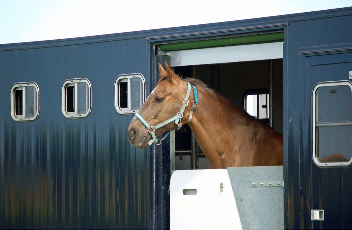
It’s a disease associated with conditions during travel for sporting, medical, or reproductive purposes. Because these trips are often numerous when it comes to horses, the probability of developing this infection makes it important to take it into account. In the following article, we’ll tell you all about it.
Equine pleuropneumonia: Characteristics of the disease
We’ll now describe what characteristics make equine pleuropneumonia a major concern among horse owners.
Origin and causes
During routine travel, horses travel in containers in conditions that are less than ideal:
- Temperatures are often inadequate, as is ventilation.
- Hygiene inside the vehicle is often deficient.
This worsens the condition of the horse’s respiratory system, which encourages any bacteria that were previously commensal to infect the lungs. In other words, equine pleuropneumonia is the consequence of a temporary state of immunosuppression aggravated by the stress of travel.
Causative agent
By definition, this disease is an infection caused by bacteria. This is the case, for example, of Streptococcus equi, a bacterium inhabiting the pharynx that can also cause mumps. Numerous species capable of producing the syndrome known as equine pleuropneumonia have been discovered.
As its name suggests, it’s a disease that affects the lungs and pleural space. In other words, it directly damages the organs responsible for respiration. Therefore, its consequences are very serious.
The evolution of equine pleuropneumonia
The disease usually develops in three consecutive phases:
- It starts with an exudative phase, i.e. with mucus due to local inflammation. If appropriate treatment is given at this early stage, the disease may not progress.
- If it does progress, the fibropurulent phase will begin. This involves the accumulation of pus and fibrin, which are nothing more than the remains of the animal’s inflammatory response.
- The last phase is the reorganization of the immune system. This generates a sort of membrane that covers the lung in order to prevent the spread of the bacteria to the rest of the body. Unfortunately, this only worsens the respiratory distress of the horse.
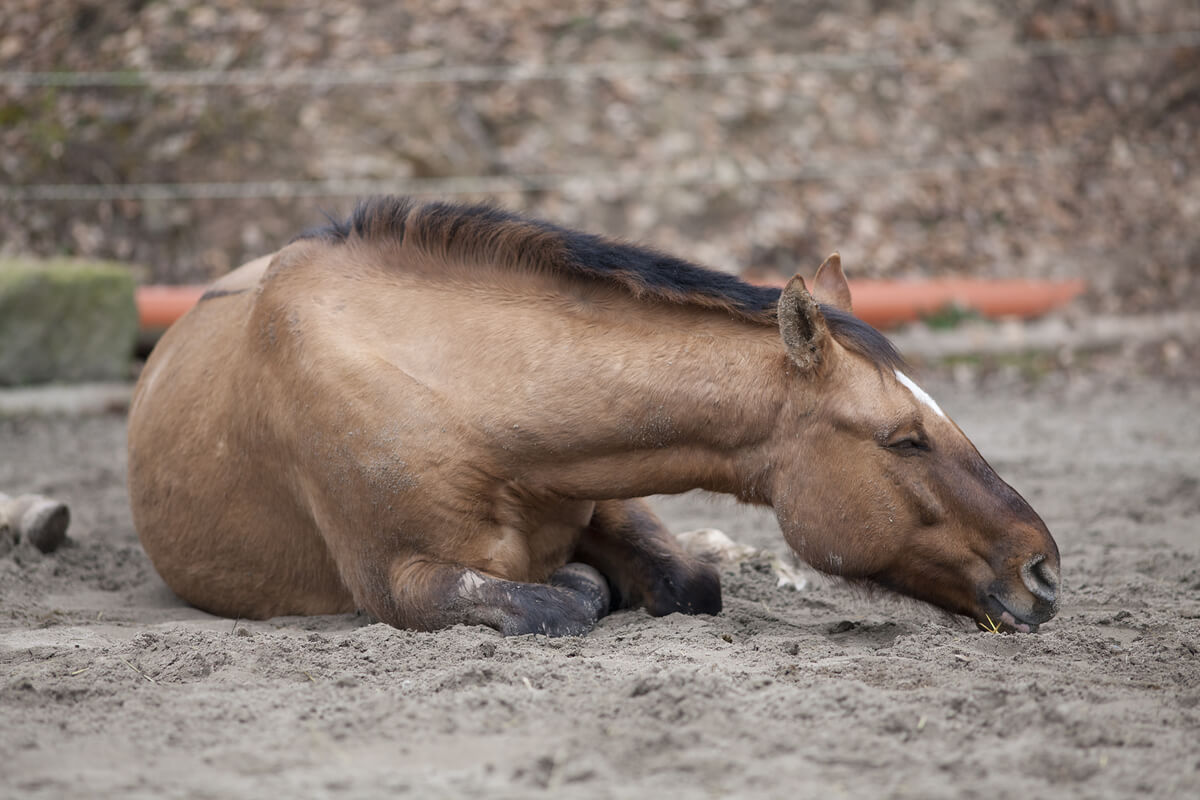
Symptoms
- Fever.
- Lethargy and depression.
- Cough, respiratory distress, and nasal mucus.
- Exercise intolerance. This refers to fatigue and the impossibility of continuing with the activity due to respiratory insufficiency.
- Inappetence.
- Often, there’s an anxious facial expression due to the horse’s “pleural pain.” Also, the animal’s movements become stiff due to pain.
In chronic cases, the fever is usually intermittent, and what’s detected is a gradual worsening of the animal’s health. While the external symptoms appear less severe or obvious, disaster is looming inside the body. Most likely, the horse will eventually suffer from sepsis, which will lead to illness and death.
The diagnosis of equine pleuropneumonia
Because the symptoms are confusing, as they’re shared with a multitude of other diseases, other diagnostic tests will be necessary. The most useful to date has proven to be ultrasound of the thoracic region. The use of ultrasound can reveal abnormal accumulation of fluid in the pleural cavity, changes in lung tissue, and the formation of abscesses.
Another useful tool is thoracentesis, which is the surgical puncture of the chest wall. This makes it possible to collect a sample of the pleural contents and identify its characteristics. In addition, this can greatly relieve pressure within the thorax, which facilitates breathing.
Tracheal aspiration can also be effective. This technique allows the aspirated fluid to be cultured in order to attempt to determine the causative agent of the infection. Subsequent treatment will then be directed against the bacteria in question.
Treatment
Because the causative agents are bacterial, treatment will focus on the administration of antibiotics. In addition, this therapy should probably be accompanied by anti-inflammatory and analgesic drugs in order to reduce the inflammation gradually.
To improve respiratory symptoms, drainage of the pleural fluid may be an option. This way, the lungs will be able to expand normally again, occupying all the space that they should.
In those cases in which breathing is really compromised, oxygen therapy and bronchodilators should be considered. But be careful, as bronchodilators can leave residue for some time, and this can trigger doping problems.
The prognosis of equine pleuropneumonia
The prognosis is usually favorable, at least in cases where the disease is identified early on, and horses receive prompt aggressive treatment. In contrast, chronic cases tend to get much worse and become complicated by laminitis, colic, and other processes.
In any case, the possible sequelae of this disease are varied and include the following:
- Pulmonary abscesses: Cavities that form within the lung tissue and fill with pus, a process that destroys respiratory cells.
- Bronchopleural fistulas: Abnormal communications between the bronchi and the pleural space.
- Pneumothorax: The entry of air into the thorax through a wound on the surface of the ribs, which compresses the lungs and prevents breathing.
- Restrictive pericarditis: The inflammation of the pericardium that surrounds the heart, preventing it from pumping blood normally.
As you can see, these are serious complications that can jeopardize the animal’s survival or at least its productive capacity as a competition or traction horse. It’s very important to protect equines against these diseases, as the outcome can be fatal.
Equine pleuropneumonia occurs due to the colonization of the animal’s lung tissue by different bacteria. These microorganisms don’t limit themselves to damage the organ but spread to the pleura that covers it and to the virtual space that separates it from the rest. Its occurrence is associated with the previous presence of bacteria in the pharynx due to catarrh or similar diseases.
It’s a disease associated with conditions during travel for sporting, medical, or reproductive purposes. Because these trips are often numerous when it comes to horses, the probability of developing this infection makes it important to take it into account. In the following article, we’ll tell you all about it.
Equine pleuropneumonia: Characteristics of the disease
We’ll now describe what characteristics make equine pleuropneumonia a major concern among horse owners.
Origin and causes
During routine travel, horses travel in containers in conditions that are less than ideal:
- Temperatures are often inadequate, as is ventilation.
- Hygiene inside the vehicle is often deficient.
This worsens the condition of the horse’s respiratory system, which encourages any bacteria that were previously commensal to infect the lungs. In other words, equine pleuropneumonia is the consequence of a temporary state of immunosuppression aggravated by the stress of travel.
Causative agent
By definition, this disease is an infection caused by bacteria. This is the case, for example, of Streptococcus equi, a bacterium inhabiting the pharynx that can also cause mumps. Numerous species capable of producing the syndrome known as equine pleuropneumonia have been discovered.
As its name suggests, it’s a disease that affects the lungs and pleural space. In other words, it directly damages the organs responsible for respiration. Therefore, its consequences are very serious.
The evolution of equine pleuropneumonia
The disease usually develops in three consecutive phases:
- It starts with an exudative phase, i.e. with mucus due to local inflammation. If appropriate treatment is given at this early stage, the disease may not progress.
- If it does progress, the fibropurulent phase will begin. This involves the accumulation of pus and fibrin, which are nothing more than the remains of the animal’s inflammatory response.
- The last phase is the reorganization of the immune system. This generates a sort of membrane that covers the lung in order to prevent the spread of the bacteria to the rest of the body. Unfortunately, this only worsens the respiratory distress of the horse.
Symptoms
- Fever.
- Lethargy and depression.
- Cough, respiratory distress, and nasal mucus.
- Exercise intolerance. This refers to fatigue and the impossibility of continuing with the activity due to respiratory insufficiency.
- Inappetence.
- Often, there’s an anxious facial expression due to the horse’s “pleural pain.” Also, the animal’s movements become stiff due to pain.
In chronic cases, the fever is usually intermittent, and what’s detected is a gradual worsening of the animal’s health. While the external symptoms appear less severe or obvious, disaster is looming inside the body. Most likely, the horse will eventually suffer from sepsis, which will lead to illness and death.
The diagnosis of equine pleuropneumonia
Because the symptoms are confusing, as they’re shared with a multitude of other diseases, other diagnostic tests will be necessary. The most useful to date has proven to be ultrasound of the thoracic region. The use of ultrasound can reveal abnormal accumulation of fluid in the pleural cavity, changes in lung tissue, and the formation of abscesses.
Another useful tool is thoracentesis, which is the surgical puncture of the chest wall. This makes it possible to collect a sample of the pleural contents and identify its characteristics. In addition, this can greatly relieve pressure within the thorax, which facilitates breathing.
Tracheal aspiration can also be effective. This technique allows the aspirated fluid to be cultured in order to attempt to determine the causative agent of the infection. Subsequent treatment will then be directed against the bacteria in question.
Treatment
Because the causative agents are bacterial, treatment will focus on the administration of antibiotics. In addition, this therapy should probably be accompanied by anti-inflammatory and analgesic drugs in order to reduce the inflammation gradually.
To improve respiratory symptoms, drainage of the pleural fluid may be an option. This way, the lungs will be able to expand normally again, occupying all the space that they should.
In those cases in which breathing is really compromised, oxygen therapy and bronchodilators should be considered. But be careful, as bronchodilators can leave residue for some time, and this can trigger doping problems.
The prognosis of equine pleuropneumonia
The prognosis is usually favorable, at least in cases where the disease is identified early on, and horses receive prompt aggressive treatment. In contrast, chronic cases tend to get much worse and become complicated by laminitis, colic, and other processes.
In any case, the possible sequelae of this disease are varied and include the following:
- Pulmonary abscesses: Cavities that form within the lung tissue and fill with pus, a process that destroys respiratory cells.
- Bronchopleural fistulas: Abnormal communications between the bronchi and the pleural space.
- Pneumothorax: The entry of air into the thorax through a wound on the surface of the ribs, which compresses the lungs and prevents breathing.
- Restrictive pericarditis: The inflammation of the pericardium that surrounds the heart, preventing it from pumping blood normally.
As you can see, these are serious complications that can jeopardize the animal’s survival or at least its productive capacity as a competition or traction horse. It’s very important to protect equines against these diseases, as the outcome can be fatal.
All cited sources were thoroughly reviewed by our team to ensure their quality, reliability, currency, and validity. The bibliography of this article was considered reliable and of academic or scientific accuracy.
- Pleura [Internet]. Es.wikipedia.org. [cited 15 September 2020]. Available from: https://es.wikipedia.org/wiki/Pleura#:~:text=La%20cavidad%20pleural%20es%20un,capa%20de%20l%C3%ADquido%20casi%20capilar.
- Comensalismo [Internet]. Es.wikipedia.org. [cited 15 September 2020]. Available from: https://es.wikipedia.org/wiki/Comensalismo
- Fibrina [Internet]. Es.wikipedia.org. [cited 15 September 2020]. Available from: https://es.wikipedia.org/wiki/Fibrina
- Aguilera Tejero E, Díez de Castro E, Mayer Valor R. Pleuroneumonía Equina (Equine Pleuropneumonia) [Internet]. AGRIS: International Information System for the Agricultural Science and Technology. 2009 [cited 15 September 2020]. Available from: https://agris.fao.org/agris-search/search.do?recordID=DJ2012040599
- Rivero L. Pleuroneumonía en equinos [Internet]. 2012 [cited 15 September 2020]. Available from: http://produccion-animal.com.ar/produccion_equinos/Enfermedades/02-pleuroneumonia-en-equinos.pdf
This text is provided for informational purposes only and does not replace consultation with a professional. If in doubt, consult your specialist.