Stomatitis in Reptiles: Causes, Symptoms and Treatment


Written and verified by the biologist Samuel Sanchez
Stomatitis is a very common condition in wild reptiles that end up in the illegal pet trade. This condition is usually derived from stress, poor care, immunosuppression of the specimen, and a poor quality of life in general. The disease that concerns us here shows that removing animals from their natural environment for personal enjoyment is never an option.
Stomatitis is more observable in snakes than in other reptile taxa, but all are susceptible to it. Read on, and find out everything you need to know about this potentially lethal infectious disease.
What is stomatitis?
Stomatitis is defined as an inflammatory response in the oral cavity of the reptile to a traumatic, infectious, metabolic or neoplastic (tumor) condition. This is one of the many conditions included in the group of upper alimentary tract diseases (UATD), along with esophageal, pharyngeal, and dental pathologies.
Stomatitis mostly affects ophidians (snakes) that have been removed from their natural habitat to be kept in captivity. Depending on its severity, it’s divided into the following stages:
- Phase I (acute): There’s an increase in the density of saliva secreted by the affected animal. Also characteristic of this stage are petechiae (red spots) in the oral cavity.
- Phase II (purulent): As the name suggests, in this phase, the reptile begins to produce pus in the oral cavity. Purulent plaques are formed and, in the most severe cases, facial deformation occurs.
- Phase III (tooth loss): Necrosis of the gingival tissue (gums) occurs and the animal’s teeth fall out. In some cases, the tongue may also become completely detached.
If left untreated, this clinical entity can lead to osteomyelitis (bone infection), pneumonia, and infectious gastritis. In other words, the bacteria that infect the mouth are capable of spreading to the rest of the animal’s organs and causing severe systemic involvement.
Causes
The causes of stomatitis are very diverse, although they all have to do with poor management and poor care of the reptile in captivity. We can divide its etiology into 2 groups, but keep in mind that both are related to each other.
Infectious causes
Infectious causes trigger stomatitis in all cases. This clinical condition may result from an overgrowth of commensal bacteria within the oral environment of the reptile. Some of the most commonly reported bacterial genera are Aeromonas spp. Pseudomonas spp., Klebsiella spp. Proteus spp . and Salmonella spp.
Some viral agents also trigger stomatitis, especially in tortoises. The most common viruses are Paramyxoviridae, Retroviridae and Herpesviridae. These viruses are usually permanent, i.e. the infection reactivates or decreases in intensity depending on the reptile’s health, but never disappears completely.
Some complex organs and parasites can cause stomatitis, but these are rare.
Handling and physical causes
This category includes all triggers that may cause physiological stress in the animal. A prolonged state of alertness results in immunosuppression, or in other words immune deficiency. Therefore, the reptile will be more prone to infection by organisms that in a normal situation aren’t harmful.
Excessive terrarium humidity, inadequate substrate, too low a temperature, lack of environmental enrichment, cramped space and malnutrition are the main causative agents in this category. Vitamin C deficiency and calcium/phosphorus imbalances have been identified in many cases.
Sometimes the disease is also associated with physical injury to the mouth caused by prey during hunting.
Symptoms of stomatitis in reptiles
Symptoms of stomatitis in reptiles vary according to the progression of the infection. However, there are several general clinical signs that are easy to identify.
- Anorexia. As you can imagine, a reptile with stomatitis will stop eating in all cases, especially in stages II and III of the disease.
- Dysphagia or trouble swallowing prey. This clinical sign is especially obvious in snakes, as they often unhinge their jaws to swallow very large prey.
- Excessive salivation in phase I.
- Excretion of pus from the mouth and reddening of the mouth in stages II and III.
- Cranial malformations, the inability to close their mouths, and a loss of teeth in severe cases.
- Systemic symptoms of pneumonia and gastritis if the infection spreads throughout the body.
The most common clinical sign is the presence of a whitish film around the reptile’s mouth. It’s very important to go to the vet as soon as this occurs, as the infection can quickly spread to the lungs and digestive tract. At this point, general respiratory and gastric symptoms appear.
Diagnosis
Any diagnosis begins with the vet asking the owner questions, as they’ll need to detect any possible negligence on the part of the owner. They’ll need to be honest and describe the conditions of the terrarium and the food they give to the animal. If not, it may take far too long to detect the exact problem.
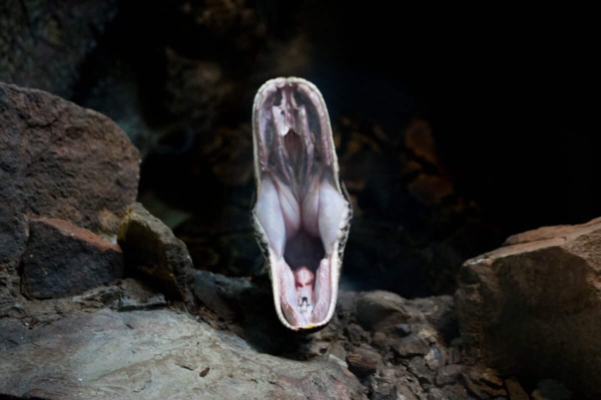
As professional sources indicate, the vet will need to open the reptile’s oral cavity with special instruments to detect possible damage to the mucous membranes. One of the most important parameters to detect is the presence of secretions, the appearance of ulcers, and whether or not the glottis can open and close correctly.
In addition to the physical examination, they’ll need to perform a blood test, a microbial culture of the oral mucosa, and an analysis of the oral tissue under a microscope. Radiographs are only necessary when it’s suspected that an abscess has caused damage to the animal’s bones.
Treatment
Treatment is always aimed at preventing the spread of the pathogen and keeping the animal metabolically active (eating and drinking). First of all, the reptile will need oral cleansing with 1% iodine and 0.25% chlorhexidine solutions every day, either in the clinic or at home.
In mild and moderate cases, local antibiotics are used, especially tetracyclines, quinolones, and aminoglycosides. However, in severe cases, the medication should be administered systemically, as the bacteria will have colonized other parts of the reptile’s body. Remember that these drugs aren’t of any use in viral and fungal infections.
If the animal arrives at the clinic anorexic or dehydrated, it’ll need the administration of support serum until it recovers its normal metabolic levels. It’s very important to avoid dehydration, as this will only increase the likelihood of death.
Once stabilized, it’s necessary to check the general condition of the reptile’s terrarium and correct erroneous parameters. The diet should also be checked.
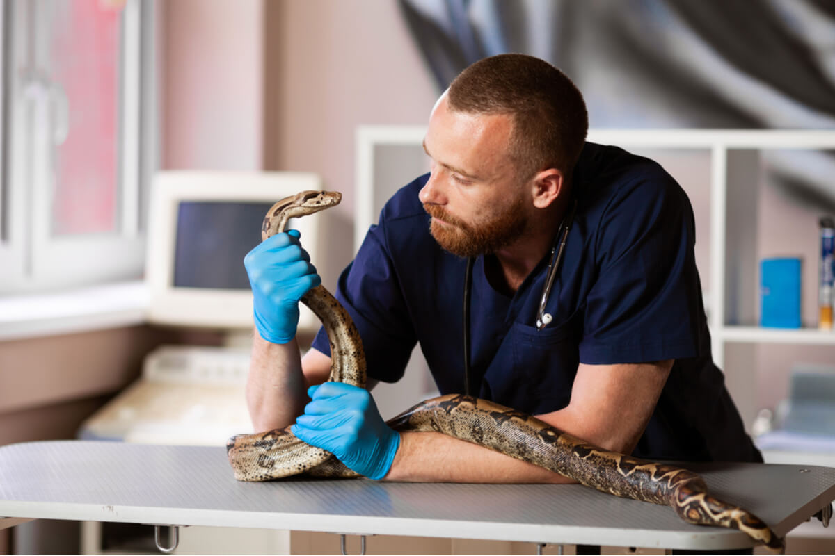
The prognosis of the condition is very dependent on the commitment of the owner. This condition can be cured, but the disease will return if the reptile’s conditions remain the same. The owners need to check the requirements for each individual species of exotic animal and follow them to the letter in order to avoid this type of potentially fatal ailment.
Stomatitis is a very common condition in wild reptiles that end up in the illegal pet trade. This condition is usually derived from stress, poor care, immunosuppression of the specimen, and a poor quality of life in general. The disease that concerns us here shows that removing animals from their natural environment for personal enjoyment is never an option.
Stomatitis is more observable in snakes than in other reptile taxa, but all are susceptible to it. Read on, and find out everything you need to know about this potentially lethal infectious disease.
What is stomatitis?
Stomatitis is defined as an inflammatory response in the oral cavity of the reptile to a traumatic, infectious, metabolic or neoplastic (tumor) condition. This is one of the many conditions included in the group of upper alimentary tract diseases (UATD), along with esophageal, pharyngeal, and dental pathologies.
Stomatitis mostly affects ophidians (snakes) that have been removed from their natural habitat to be kept in captivity. Depending on its severity, it’s divided into the following stages:
- Phase I (acute): There’s an increase in the density of saliva secreted by the affected animal. Also characteristic of this stage are petechiae (red spots) in the oral cavity.
- Phase II (purulent): As the name suggests, in this phase, the reptile begins to produce pus in the oral cavity. Purulent plaques are formed and, in the most severe cases, facial deformation occurs.
- Phase III (tooth loss): Necrosis of the gingival tissue (gums) occurs and the animal’s teeth fall out. In some cases, the tongue may also become completely detached.
If left untreated, this clinical entity can lead to osteomyelitis (bone infection), pneumonia, and infectious gastritis. In other words, the bacteria that infect the mouth are capable of spreading to the rest of the animal’s organs and causing severe systemic involvement.

Causes
The causes of stomatitis are very diverse, although they all have to do with poor management and poor care of the reptile in captivity. We can divide its etiology into 2 groups, but keep in mind that both are related to each other.
Infectious causes
Infectious causes trigger stomatitis in all cases. This clinical condition may result from an overgrowth of commensal bacteria within the oral environment of the reptile. Some of the most commonly reported bacterial genera are Aeromonas spp. Pseudomonas spp., Klebsiella spp. Proteus spp . and Salmonella spp.
Some viral agents also trigger stomatitis, especially in tortoises. The most common viruses are Paramyxoviridae, Retroviridae and Herpesviridae. These viruses are usually permanent, i.e. the infection reactivates or decreases in intensity depending on the reptile’s health, but never disappears completely.
Some complex organs and parasites can cause stomatitis, but these are rare.
Handling and physical causes
This category includes all triggers that may cause physiological stress in the animal. A prolonged state of alertness results in immunosuppression, or in other words immune deficiency. Therefore, the reptile will be more prone to infection by organisms that in a normal situation aren’t harmful.
Excessive terrarium humidity, inadequate substrate, too low a temperature, lack of environmental enrichment, cramped space and malnutrition are the main causative agents in this category. Vitamin C deficiency and calcium/phosphorus imbalances have been identified in many cases.
Sometimes the disease is also associated with physical injury to the mouth caused by prey during hunting.
Symptoms of stomatitis in reptiles
Symptoms of stomatitis in reptiles vary according to the progression of the infection. However, there are several general clinical signs that are easy to identify.
- Anorexia. As you can imagine, a reptile with stomatitis will stop eating in all cases, especially in stages II and III of the disease.
- Dysphagia or trouble swallowing prey. This clinical sign is especially obvious in snakes, as they often unhinge their jaws to swallow very large prey.
- Excessive salivation in phase I.
- Excretion of pus from the mouth and reddening of the mouth in stages II and III.
- Cranial malformations, the inability to close their mouths, and a loss of teeth in severe cases.
- Systemic symptoms of pneumonia and gastritis if the infection spreads throughout the body.
The most common clinical sign is the presence of a whitish film around the reptile’s mouth. It’s very important to go to the vet as soon as this occurs, as the infection can quickly spread to the lungs and digestive tract. At this point, general respiratory and gastric symptoms appear.
Diagnosis
Any diagnosis begins with the vet asking the owner questions, as they’ll need to detect any possible negligence on the part of the owner. They’ll need to be honest and describe the conditions of the terrarium and the food they give to the animal. If not, it may take far too long to detect the exact problem.
As professional sources indicate, the vet will need to open the reptile’s oral cavity with special instruments to detect possible damage to the mucous membranes. One of the most important parameters to detect is the presence of secretions, the appearance of ulcers, and whether or not the glottis can open and close correctly.
In addition to the physical examination, they’ll need to perform a blood test, a microbial culture of the oral mucosa, and an analysis of the oral tissue under a microscope. Radiographs are only necessary when it’s suspected that an abscess has caused damage to the animal’s bones.
Treatment
Treatment is always aimed at preventing the spread of the pathogen and keeping the animal metabolically active (eating and drinking). First of all, the reptile will need oral cleansing with 1% iodine and 0.25% chlorhexidine solutions every day, either in the clinic or at home.
In mild and moderate cases, local antibiotics are used, especially tetracyclines, quinolones, and aminoglycosides. However, in severe cases, the medication should be administered systemically, as the bacteria will have colonized other parts of the reptile’s body. Remember that these drugs aren’t of any use in viral and fungal infections.
If the animal arrives at the clinic anorexic or dehydrated, it’ll need the administration of support serum until it recovers its normal metabolic levels. It’s very important to avoid dehydration, as this will only increase the likelihood of death.
Once stabilized, it’s necessary to check the general condition of the reptile’s terrarium and correct erroneous parameters. The diet should also be checked.

The prognosis of the condition is very dependent on the commitment of the owner. This condition can be cured, but the disease will return if the reptile’s conditions remain the same. The owners need to check the requirements for each individual species of exotic animal and follow them to the letter in order to avoid this type of potentially fatal ailment.
All cited sources were thoroughly reviewed by our team to ensure their quality, reliability, currency, and validity. The bibliography of this article was considered reliable and of academic or scientific accuracy.
- Revisión de la estomatitis ulcerativa en reptiles, PDF. Recogido a 21 de octubre en https://www.redalyc.org/pdf/4076/407642324007.pdf
- Infectious Stomatitis (Mouth Rot) in Reptiles, PetCoach. Recogido a 21 de octubre en https://www.petcoach.co/article/infectious-stomatitis-mouth-rot-in-reptiles-causes-signs-di/
- Lizard and Snake Ulcerative Stomatitis, WikiVet. Recogido a 21 de octubre en https://en.wikivet.net/Lizard_and_Snake_Ulcerative_Stomatitis
This text is provided for informational purposes only and does not replace consultation with a professional. If in doubt, consult your specialist.


